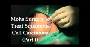
Mohs Surgery to Treat Squamous Cell Carcinoma
Mohs Surgery to Treat Squamous Cell Carcinoma
 In this multi-part video series, Dr. Evans Bailey walks us through a Mohs surgery procedure to treat squamous cell carcinoma. If left untreated, squamous cell carcinoma carries the risk of spreading to the lymph nodes, making it exceptionally important to address.
As an elected member of the American College of Mohs Surgery and fellowship-trained skin cancer and reconstructive surgeon, Dr. Bailey acts as both the surgeon and pathologist in the tissue-sparing Mohs surgical procedure. Named for Dr. Fred Mohs, the Mohs surgery technique treats skin cancer by gradually removing thin layers of skin from a skin cancer site until a patient is cancer free. At the time of surgery, Dr. Bailey is able to process the skin tissue, evaluate it and take more if necessary to ensure no cancerous tissue remains around the surgical site. According to Dr. Bailey, “That’s really what the Mohs technique involves, removing the tumor with a small safety margin and then checking it comprehensively.”
To begin the procedure, Dr. Bailey numbs the tumor area, located on the patient’s cheek. As seen in the video, the visible tumor is the inner circle in the center, an area of invasive squamous cell. Many times these cancers will start as a scaly spot and quickly grow to a much larger invasive area.
Dr. Bailey begins the procedure by debulking the cancer, removing the top of it to allow access to the deep part of the invasive tumor. Using an instrument called a curette, he removes the bulk of the fragile tumor. He starts with a small and controlled margin, the dotted circle around the tumor. The goal is to take the minimum margin necessary, while also ensuring that the tumor area is clear. Upon removal, he processes the tissue and evaluates it in 3 dimensions, allowing for a precise analysis under the microscope. Mohs surgery, the most accurate technique to make sure the cancer is fully removed, allows for Dr. Bailey to assess if the tumor is invading any part beyond what he has removed through a comprehensive margin assessment.
Stay tuned for our next post in this series of Mohs surgery videos. As you’ll see in Part Three, Dr. Bailey allows us inside the lab at Naaman Clinic to observe the processing of the tissue.
