Mohs Surgery to Treat Squamous Cell Carcinoma Part Two
 As seen in Part One of this multi-part video series, Dr. Bailey performed a Mohs surgery procedure to treat squamous cell carcinoma. In this specific case, the tumor was located on the patient’s cheek and after removing the necessary margin, Dr. Bailey is ready to begin the precise analysis of the tissue in the lab at Naaman Clinic.
You’ll recall that Mohs surgery, the most accurate technique to make sure the cancer is fully removed, allows Dr. Bailey to determine if the tumor is invading any area beyond what he has removed through a comprehensive margin assessment.
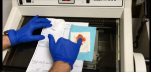
In the lab, Dr. Bailey introduces us to the cryostat, the instrument he uses to process the tissue. Mapping of the tissue and careful observation under the microscope allows for identification of tumor anywhere in the perimeter or on the underside. Dr. Bailey processes the tumor by taking thin stair-step sections from the outside edge to the center of the tissue. If there is tumor at the margin, he knows exactly where to surgically remove more tissue.
In our final video in this series, the slides are ready and Dr. Bailey studies them in the lab to see if the margins are clear. This process allows for a 3-dimensional evaluation of the specimen to see if the tumor is fully removed. As explained by Dr. Bailey, “If there is tumor anywhere on this I can see it, mark it on the map, then go back to the patient and take another sample just specifically where the tumor is. The Mohs surgery procedure ensures we are doing this with the highest cure rate but only taking what we need to.”
Going from the very edge to the center of the specimen, Dr. Bailey confirms the perimeter is completely clear. Therefore, the patient is clear of the squamous cell carcinoma and he can then reconstruct the patient.